목차
1. 개요
이번 글에서는 ankle ligament에 대해서 공부하였습니다. 아래 그림에서처럼 Lateral collateral ligament complex, Medial collateral ligament complex, Distal tibiofibular syndesmosis 순으로 정리하였습니다.


2. Lateral collateral ligaments
ATFL, CFL, PTFL 로 구성된다.
2-1 . ATFL(anterior talofibular ligament)
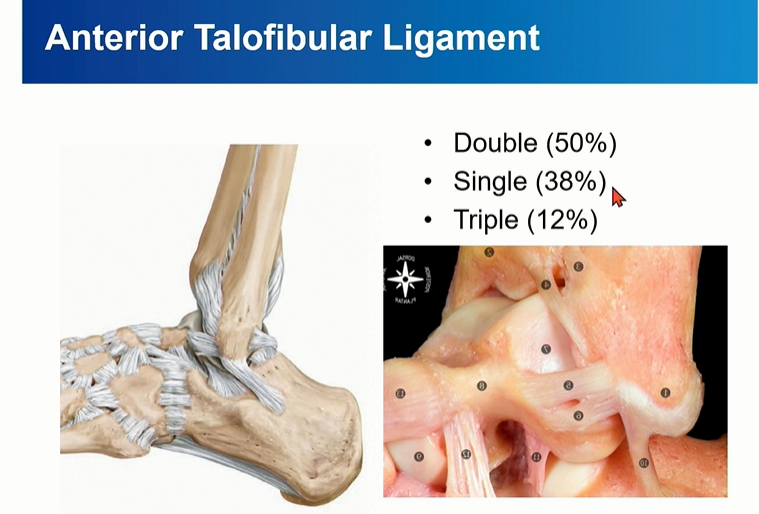
Double bundle로 되어 있는 경우가 있음을 염두.
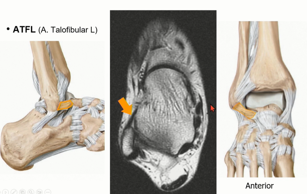
ATFL은 Axial cut에서 잘 관찰되며, fibula가 강낭콩 모양인 레벨에서 앞으로 주행하는 것을 찾으면 된다.
(그것 보다 윗 레벨에서는 joint capsule이나 AITFL을 ATFL로 오인할 수 있다)
2-2 . CFL (Calcaneofibular ligament)

ATFL과 떨어져서 기시한다는 이야기도 있었지만, Just below ATFL로 이해하면 좋을 것 같다.
Peroneal tendon을 landmark로 이용해서 CFL를 찾는 것도 연습해야 함.


Axial cut과 coronal cut에서 모두 확인할 수 있는데, Axial cut의 경우에는 peroneal tendon 내부에 위치한 것을 확인할 수 있다.
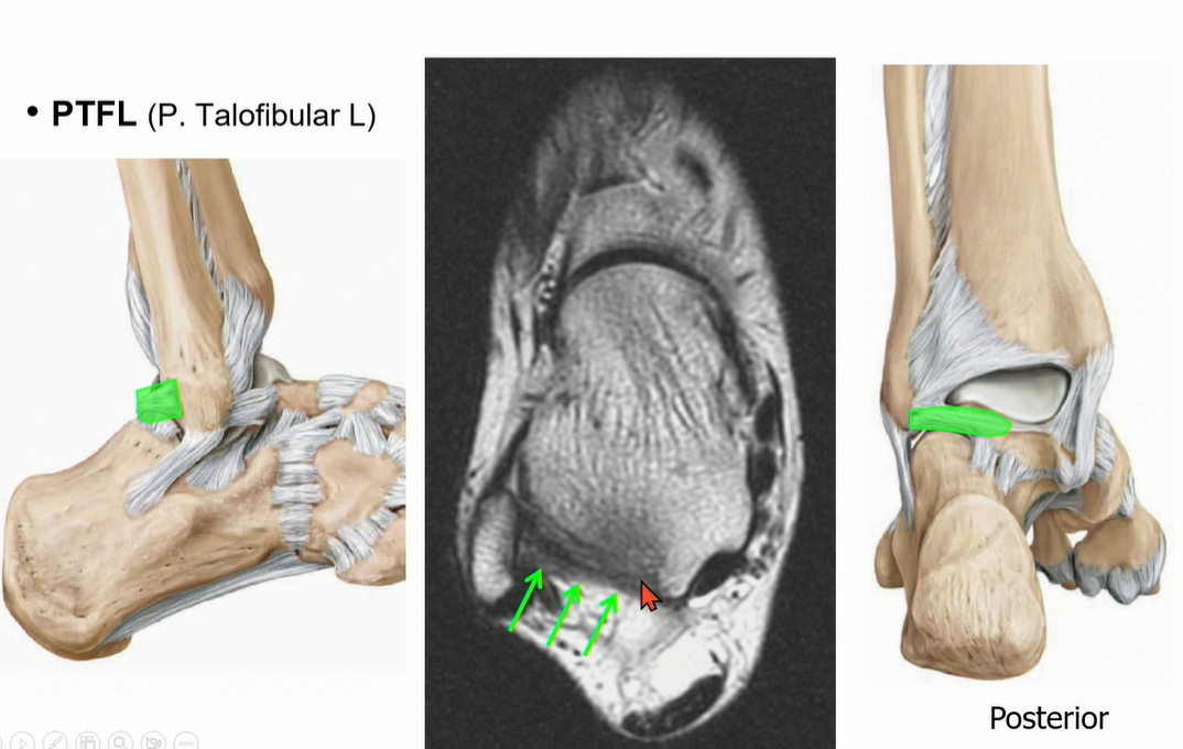
2-3 . PTFL (posterior talofibular ligament)

3가지 중에서 가장 두껍고 튼튼해서 injury가 제일 적다. 정상적인 상태에서도 ligament 내부로 fat portion이 있음.
3. Deltoid ligament

Superfical Deltoid ligament: 1-3
Deep Deltoid ligament: 4-5
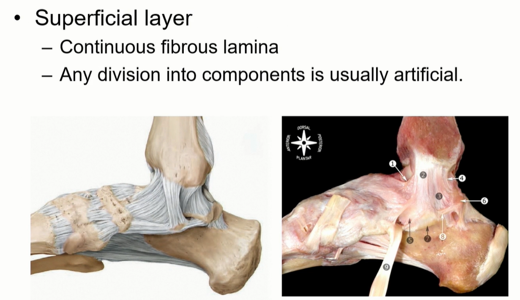
Superficial은 사실상 연속적인 구조로 되어 있다. 구분은 인위적.
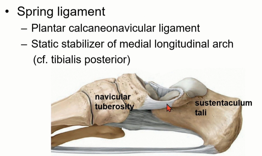
스프링 ligament는 plantar calcaneonavicular ligament라고 부르고, navicular tuberosity와 sustentaculum tali를 이어주는 인대이다. Medial longitudinal arch의 static stabilizer 중에 하나임. (다른 하나는 TP tendon )
-> Flat foot의 경우에는 2개의 static stabilizer 평가가 중요하다.

Spring ligament는 1-3으로 구성되어 있다. complex임.
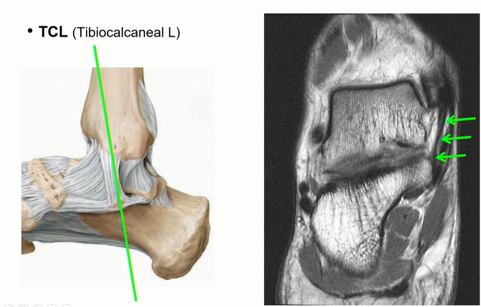
Tibiocalcaneal ligament는 calcaneus의 sustentaculum tali로 이어짐. 비교적 수직으로 주행하기 때문에 관찰 용이.
(Deltoid ligament의 superficial ligament의 3번에 해당)

Tibiospring ligament는 Deltoid ligament의 superficial ligament의 2번에 해당.
spring ligament에 붙는다.

Deltoid ligament의 superficial ligament의 1번에 해당하는 tibionavicular ligement는 주행이 비스듬해서 coronal에서는 잘 관찰이 어렵다. (navicular tuberosity에 붙음)

Deep deltoid liagment의 하나인 PTTL의 주행.
ATTL에 비해서 넓어서 영상에서 확인이 용이하다. ligament 사이에 fat이 관찰되는 것이 정상.
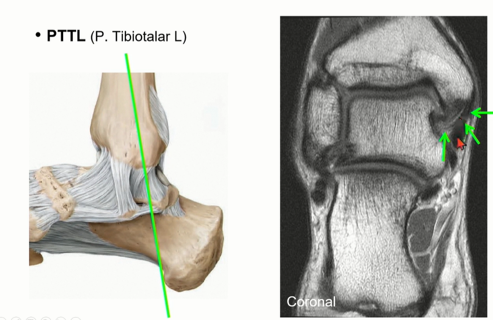
coronal cut에서도 fat때문에 straiation이 보이는 것이 정상이다.

녹색으로 표시한 것은 flexor retinaculum으로 deltoid ligament와 오인하지 않도록 조심해야 한다.
4. Distal tibiofibular syndemosis
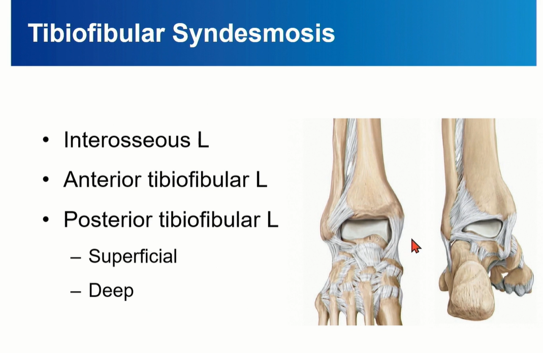
Tibiofibular syndemosis = Interosseous Ligament + AITFL + PITFL
PITFL 은 superfical component와 deep component로 구성되는데, deep component를 inferior transverse ligament라고 부름.
4-1. Interosseous Ligament
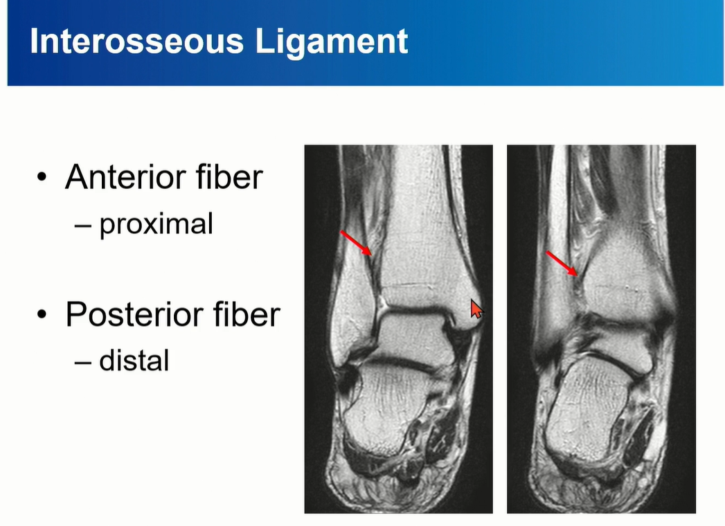
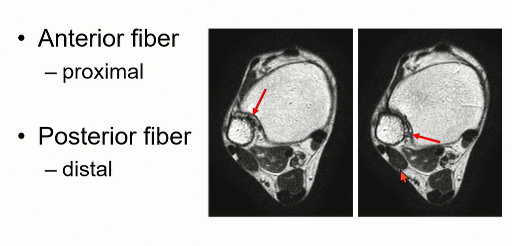
Anterior fiber가 proximal에 위치, posterior fiber가 distal에 위치하게 된다.
Tibia의 fibular notch와 fibula를 연결하는 역할을 한다.
4-2. ATTFL


Tibia plafond의 anterolateral tubercle에서 시작해서 distal fibula에 붙고, 비스듬하게 여러 가닥으로 구성되어 있다.
AITFL의 가장 inferior bundle은 main bundle에서 떨어져 있는데, 이 fiber를 Bassett's ligament 라고 부른다. Talus와 impinge를 일으키기도 하는데, anterolateral ankle impingement를 뜻함.
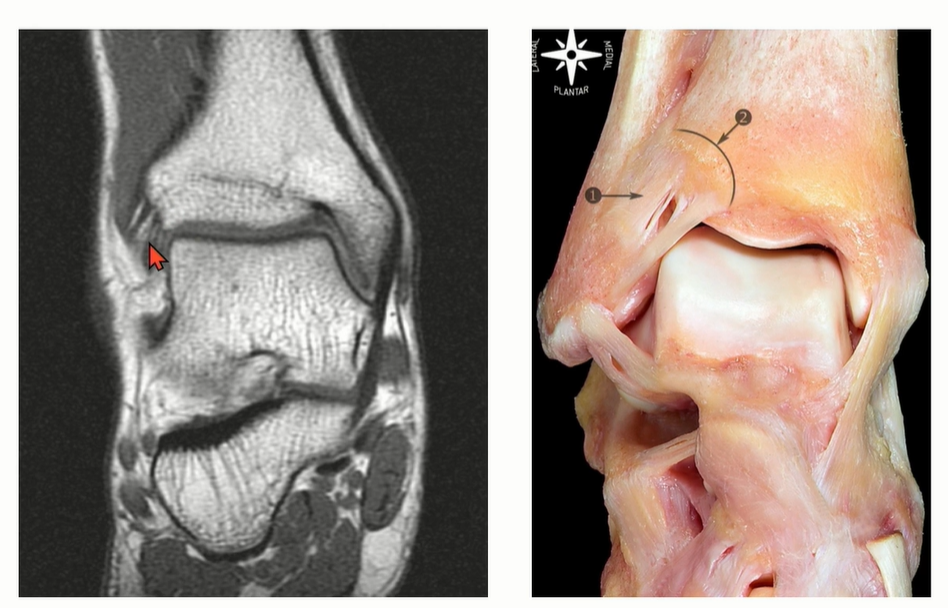
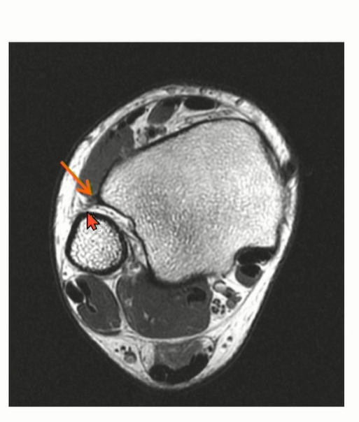
MRI에서는 coronal cut에서 AITFL 를 관찰하기 용이하다. Multi-fascicular 구조이기 떄문에, Tear로 오인하면 안됨.
특히 Axial cut에서는 일부 cut에서 discontinuity 가 관찰될 수 있는데, oblique한 주행 방향으로 인한 정상 소견일 수 있다는 것을 염두해야 한다.
4-3. PITFL
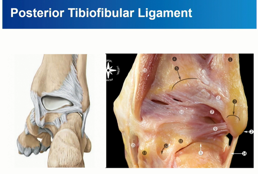
PITFL 은 superfical component와 deep component로 구성된다.
deep component를 inferior transverse ligament라고 부름. 이름대로 horizontal 하게 주행하는 것을 확인할 수 있다.

PITFL은 superficial 과 Deep으로 나누어 보기도 한다.
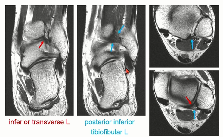
빨간색으로 표시된 inferior transverse ligament가 아래 쪽에서 주행하는 것을 확인할 수 있다.
파란색으로 표시한 posterior inferior tibiofibular ligment는 위쪽에서 oblique하게 주행하는 것을 확인할 수 있다.

Deep PITFL인 interior transverse ligament는 labrum으로 역할을 한다. 관절면을 deepening 해줌.
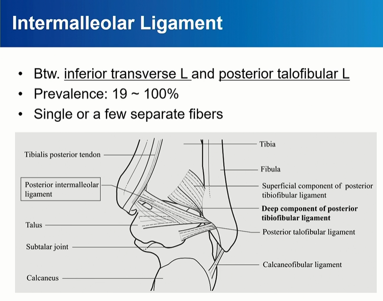
intermalleolar ligament는 PTFL 과 Deep PITFL (Inferior transverse ligament)사이에 존재.
비스듬한 주행, Lateral쪽으로 가면서 아래로 주행.
'정형외과 > 근골격 영상 공부' 카테고리의 다른 글
| 비구 골절_acetabulum fracture (4) | 2024.11.20 |
|---|---|
| 비구 골절_acetabulum fracture (1) | 2024.11.19 |
| 골반환 손상_Pelvic ring injury (0) | 2024.11.16 |
| 족부 골절 영상 공부_발목 외 기타 골절 (0) | 2024.11.14 |
| 족부 골절 영상 공부_Fracture of ankle (2) | 2024.11.13 |
| 발목 MRI 공부_Ankle instability (0) | 2024.11.12 |
| 발목 MRI 공부_Ankle impingement (0) | 2024.11.12 |
| 무릎 영상 공부(1) (0) | 2024.06.20 |
